
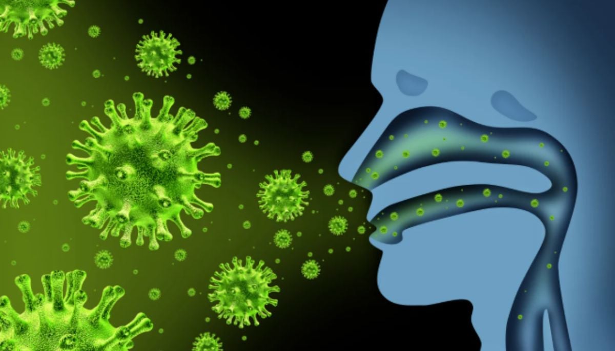
Winter has returned, and so has flu season that brings flu, fever, aching limbs and runny nose, forcing scientists to study how the virus interacts with the human body.
Scientists explained that almost all the symptoms mentioned are caused by flu viruses, which enter our body through small droplets and then infect vulnerable cells.
To study a more detailed process, a team of researchers from Switzerland and Japan took an exceptionally close look at how the virus behaves.
According to Science dailya study published in the journal Improved visualization of influenza A virus entry into living cells using atomic force microscopy with virus display, has helped scientists monitor the process using advanced techniques.
Using specialized microscopes, the scientists zoomed in on the outer surface of human cells in a petri dish to watch their activity live and examine sharp details of how the flu virus enters a living cell.
The group of researchers, together with Professor of Molecular Medicine Yohei Yamauchi at ETH Zurich, unexpectedly discovered that human cells do not enter the virus easily and make multiple attempts to grab the virus or do their utmost to prevent it from entering the body.
The membrane also pushes upward at that point, almost as if it is trying to grab the virus, and these wave-like movements become stronger as the virus tries to float away from the surface.
The study researchers showed that cells support the virus at different stages of entry.
Although cells gain nothing by being infected, the action still appears to be active, as the virus disrupts a routine cellular uptake system that the cells cannot do without.
“The infection of our body cells is like a dance between virus and cell,” says Yamauchi.
As they dug into the details, scientists noticed that the influenza virus attaches to specific molecules on the cell surface and begins to infect the body.
The process is similar to membrane surfing, and the virus moves along the surface, attaching to one molecule after another until it reaches the most suitable receptors rich in proteins.
When the cell’s receptors detect that the virus has attached itself, the membrane begins to form a small indentation at that spot.
A structural protein called ‘Clathrin’ forms and supports this deeper pocket. As the sac enlarges, it wraps around the virus and forms a vesicle, as the cell then pulls this vesicle inside, where the coat dissolves and releases the virus.
The researchers conducted the experiment using a process called virus-view dual confocal and atomic force microscopy AFM, which combines AFM with fluorescence microscopy.
They said this combined approach makes it possible to track the fine-scale movements involved when the virus enters the cell.